Sample Preparation Lab
The sample preparation laboratory at the Department of Earth and Atmospheric Sciences (EAS) at Cornell includes rock saws, jaw crusher, shatter box, powdering mills (alumina, agate, etc.), magnetic and density separation, grain size separation equipment, and multiple polishing capabilities. The lab is outfitted with fume hoods, bench space, and utilities (deionized water, natural gas, etc.), drying ovens, sieves, and glassware.
Petrographic Microscopy
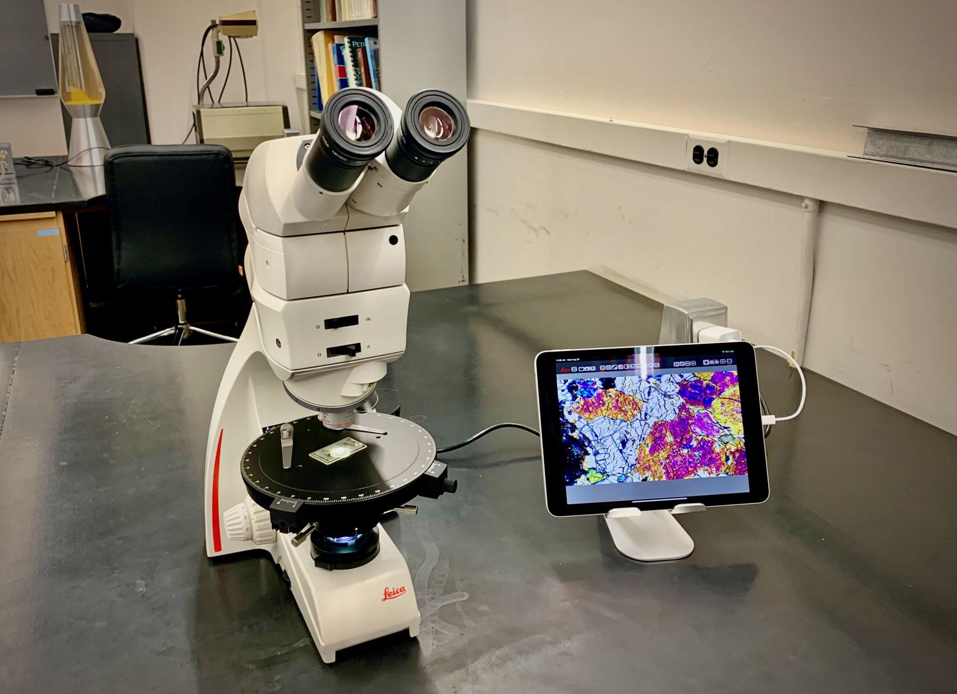
A new comprehensive microscopy laboratory includes research-grade Leica compound (petrographic) and stereographic microscopes for both transmitted and reflected light microscopy, equipped with digital photomicrography capability and dedicated computers with relevant imaging analysis software is now available at EAS.

Raman Spectroscopy
Our lab includes a new WiTec Alpha 300r Raman micro-spectrometer. This is an advanced confocal Raman microscope that combines fast imaging, high spectral sensitivity and spatial resolution with excellent analysis software into a system designed around spectral imaging. The system has 532-nm and 633-nm lasers and spectrometers covering shifts in the visible. Raman shifts within ~100 cm-1 of the excitation frequency can be measured routinely. The system has high confocality and boasts a minimum depth slice of ~350 nm, enabling 2D and 3D chemical maps of Raman intensity over areas as large as several square cm. Using 3D volume mapping of moderately absorbing samples, the Raman intensity can survey to depths of multiple microns into the sample. We also included an internal Ne source that allows to collect Ne lines simultaneously with the data, allowing a highly precise internal calibration. This instrument allows mapping at high resolution of Earth and planetary materials, while also increasing the analytical resolution for peak fitting.

Experimental Heating and Cooling Stages
- Linkam TS1500V stage with a temperature range from ambient to 1500 °C. The sample is placed inside the ceramic sample container, and it is heated uniformly from the sides and below at a rate of 200°C/min. This stage allows vacuum connectors and the use of inert gases (e.g., He, Ar) in a constant flow or after vacuum. Both reflection and transmission (e.g., for FTIR) in-situ measurements are possible with this during heating experiments.
- Linkam TS1400XY, a relatively new stage designed for melt inclusion heating and re-homogenization experiments. The temperature range goes from ambient to 1400°C at a heating rate of up to 200°C/min and cooling rates of 240°C per second. The sample is mounted onto a sapphire sample slide and can be manipulated within the ceramic heater by 6mm in the X and Y-axis. This stage will be used to re-heat, and if necessary re homogenize, melt inclusions after water data was collected by Raman spectroscopy to avoid any induced diffusion loss into the host.
- Linkam THMSG600 specifically designed for micro-thermometry of fluid inclusions with a precision better than 0.1°C and a stability of 0.001°C. The THMSG600 has a temperature range of -195°C to 600°C. The sample is placed on 7 mm quartz cover slip. A liquid nitrogen cooling pump is used to enable a wide range of cooling rates from 0.01°C to 150°C/min.
The experimental heating and cooling stages include Linkam TS96 controllers, vacuum pump, and a modified Olympus BX53MTRF-S long-working distance microscope. Specifically, for the melt inclusion experiments on olivine hosted inclusions we installed a SENTAG oxygen purge system, necessary to remove any trace O2 in the He or Ar used, and a SENTAG oxygen analyzer to control the state of oxidation of the experiments.
CO2 (and other gases) calibration apparatus
Our experimental setup at Cornell includes an optical pressure cell is a custom drilled pressure cross rated to 34.47 MPa at 40 ºC, with three high-pressure connections, one medium pressure connection for a thermal probe, and a quartz window drilled into the top. The pressure is recorded in real-time on a laptop computer at 1Hz frequency and measured directly inside the optical pressure cell using a pressure transducer with 0.15% accuracy. Temperature is also measured directly inside the optical pressure cell, using a steel probe thermometer with a resolution of 0.01ºC and an accuracy of +-0.05ºC at room temperature to 37 ºC. The optical pressure cell is connected to two standard high-pressure valves on either side. The input is connected to a manual screw press-type pressure generator, and the whole setup around the optical pressure cell is enclosed in foam to provide thermal insulation. The optical pressure cell sits on a thermoelectric Peltier plate in which the temperature can be precisely controlled and adjusted throughout the experiment. Ultra-high purity CO2 is used for the calibrations, and the optical pressure cell is purged continuously before each experiment to remove any other gases and/or water. This setup ensures that the temperature inside the optical pressure cell varies by no more than +-0.02ºC at room temperature and no more than +-0.01ºC while using the PP at 37 ºC and also ensures that the pressure inside the optical pressure cell varies by no more than +-0.004 MPa during each analysis. The positions of the Fermi diad peaks are measured in 0.5 MPa increments in the 0.05-7 MPa range at 37 ºC, in increments of 0.1 MPa in the 7-8 MPa range and 9-11, in increments of 0.05 MPa in the 8-9 MPa range and finally in 1 MPa increments in the 11-32 MPa range. This apparatus will be primarily used for CO2 but it can also be used to calibrate the Raman instrument for other gases like CH4, N, etc.

Fourier Transform Infrared Spectroscopy
We acquired a new Thermo-Nicolet iN10MX FTIR with 2D mapping capabilities using a linear array with a space resolution of ~5-10 µm, and detection limits at ppm level for water in geologic materials. This instrument allows mapping and precise measurement minerals and glasses that can be used to determine decompression rates of volcanic eruptions when combined with robust CO2 measurements determined by Raman spectroscopy.
IR to UV Spectroscopy
The Bruker Vertex 80 is equipped with detectors (MCT, DLaTGS, Si diode, GaP diode) that allow for the collection of spectra with a resolution of up to 0.06 cm-1 with a combined spectral range from the UV to the mid-IR (50000-350 cm-1; 0.2-28.6 μm). The Hyperion 2000 can do a similar range for the absorbance and transmittance of samples sitting on the microscope stage (33000-600 cm-1; 0.3-16.7 μm). Spectra can be obtained from samples inside the Vertex chamber as thin films; with an integrating sphere (8000-350 cm-1; 1.3-28.6 μm); under the Hyperion microscope 15x objective in reflection, transmission, or emission modes. We are also equipped with a Linkam heating stage to analyze material up to 1500 *C using the full spectral range of the instrument.

Mass Spectrometry
We installed at EAS an ESIL 193-nm Excimer laser with a power of up to 50 Jules connected to an Agilent 8900 triple quadrupole (LA-ICP-MS/MS) that allows to collect in-situ mineral and glass major and trace-element data. The LA-ICP-MS/MS system is dedicated for solid state samples, planned to be a regional recharge operation to service upstate NY. We also obtained in collaboration with the department of Civil and Environmental Engineering a new Agilent 7900 ICP-MS system hosted at EAS dedicated to wet chemistry trace-element analyses. Also available at EAS a Finnigan Element 2 High Resolution ICP-MS used for elemental and isotopic analyses and a SpectroBlue Inductively Coupled Plasma Optical Emission Spectrometer (ICP-OES) suited for measuring major, minor, and many trace elements in a wide variety of matrices. The ICP-OES is a complement to the ICP-MS instruments, making it easier to cover a full range of elemental abundances Earth and planetary materials.
Cornell Shared Facilities
We routinely use facilities the Scanning Electron Microscopy (SEM) and the Transmission Electron Microscopy (TEM) facilities at Cornell Center for Material Research, and the high-resolution computerized x-ray tomography at the Cornell Institute of Biotechnology (CIB). We also have access to the Cornell High Energy Synchrotron Source.